

FLASH CT
Flash CT is the world’s fastest tomography device which emits the lowest dose of radiation. It produces cardiac and lung scans in particular as well as images of almost all other parts of the body. As the device uses low doses of radiation and scans rapidly, it is instrumental, especially in cardiac check up programs. The Flash CT provides a full body tomography in just 4 seconds, a cardiac angiography in 0,25 seconds and a lung scan in 0,6 seconds without the patient having to hold his breath. The Flash CT is especially beneficial for trauma patients, pediatric patients and obese patients due to the freedom of movement during the scan and its rapidity.
PET CT
PET/CT is one of the most effective imaging techniques available today. It is particularly effective in diagnosing tumors in oncology, determining the degree to which a tumor has spread, planning radiotherapy, assessing the response to treatment and sometimes, it can determine if a mass is benign or malignant. PET/CT is the result of the combination of PET (Positron Emission Tomography) which displays the metabolic function of the organs and tissue in the human body, and CT (Computerized Tomography) which produces detailed, anatomical information. The PET/CT device is instrumental not only in oncology, but also in determining epilepsy, neurological diseases including Alzheimer’s and to pinpoint healthy tissue in the heart following a heart attack.
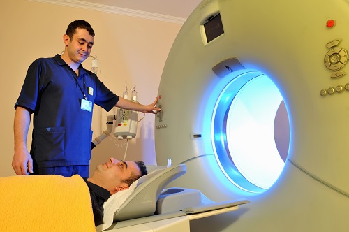
Whole Body MRI
A Whole Body MRI is relatively a new diagnostic technique which has no adverse effects. It is very effective for evaluating the entire body for patients with cancer risk or health concerns in only 30 minutes. However, with the traditional MRI, scanning only one organ takes around the same time. With the Whole Body MRI, tumors can be diagnosed in the very early stages even before the symptoms occur when the chance of treatment is at most. It can be positioned not only as an advanced diagnostic technique for diseases but also as a check-up method. During the Whole Body MRI, patient does not receive any radiation. Therefore, it is safe for every age group and can be repeated if necessary.
Intraoperative 3 Tesla MRI
Intraoperative 3 Tesla MRI is an advanced technology imaging device. It can be used to produce images of the entire body and its rapid imaging during brain surgery assists the surgeon to remove the tumor in its entirety. As the device is able to produce images during surgery, surgeons are able to carry out assessments in a sterile environment when necessary without the need to resuscitate the patient or close the surgical wound. This means that the surgeon will be able to have a more concise idea regarding the size and location of the tumor, even in places which are not easy to reach. As risks including partial removal of tumors, damage in the functional fields, operating on an incorrect area are eliminated there is no further need for further surgery.
Digital Mammography With Tomosynthesis
This is a mammography device which produces perfect two and three dimensional images by using minimal doses. This device is set to revolutionize the early diagnosis of cancer with its capacity of two dimensional and multi slice three dimensional tomosynthesis imaging. The tomosynthesis slices increase the reliability of diagnosis. A series of sliced digital images of the breast are produced which results in a three dimensional image of the breast. Routine mammography equipment displays tissue problems on one image whereas this device enables slices of the breast to be viewed digitally. This feature ensures that problem regions and breast cancer are not missed.
Single Dose Therapy
The era of single-dose radiotherapy during surgery begins…
What is Single-Dose Therapy?
In patients with breast cancer, the radiation therapy can be completed with a single-dose application done using the Intra Operative Radiotherapy (IORT) method during surgery. Once the tumor in the breast is removed in an operation, the special applicator in the tool is placed in the area with the tumor. Following the necessary calculations, high-dose radiation is given only to this reason, completing all local treatments and protecting healthy tissue.
How is Traditional Radiotherapy Applied?
Radiation therapy is carried out with “linear accelerators” that produce photons and electrons, in specially protected rooms in hospitals’ Radiation Oncology departments. In almost all cases that have been diagnosed with breast cancer and treated with “protective surgery” (in which only the portion of the breast with the tumor is removed and the rest of the breast is left intact), radiotherapy is needed following the operation.
Radiotherapy begins preferably after chemotherapy in patients who require it or at least 3 weeks after surgery in patients who are only treated with hormone therapy. In patients who’ll receive radiotherapy for the breast, tomography cross-sections with 2-mm increments are first taken using a computer for planning purposes. Later, arrangements are made to apply the necessary dose to the areas under risk in the breast that’ll receive radiotherapy while protecting the healthy organs (the heart, main arteries of the heart, lungs, other breast, etc. where the illness hasn’t spread). Once the plan is approved, the patient receives radiation treatment for 5 to 7 weeks, dependent on the condition of the illness and determined by the radiation oncology doctor. Other patients still receive a short-term treatment called hypofraction, which lasts 3 weeks.
Advantages of Single-Dose Application
The single-dose application done with the Intraoperative Radiation Therapy (IORT) tool utilizes a linear accelerator in much the same way that traditional radiotherapy does. This method is applied in an operating room using a safe and portable tool that operates in a linear and accelerated manner, only producing electron energy but not requiring too much protection by the personnel like traditional linear accelerators do. In addition to the surgical team carrying out the operation, a radiation oncology specialist and a medical physicist are also present in the operating room. This way, radiotherapy is completed at the same time as the surgery. A significant advantage for the patient is that there is no need to wait for the wound to heal following surgery, or to plan radiotherapy and visit the hospital each day for treatment. This way, the total duration of the treatment is also cut down.
During the operation, once the tumor in the breast is removed, the special applicator within the tool is placed in the area where the tumor is. Following the necessary calculations, the high-dose radiation is given only to this area, completely protecting the healthy tissue. Although single-dose application or using different methods to apply radiation only to the area with the tumor as opposed to the entire breast are methods that have become more globally accepted in recent years, single-dose application isn’t for every patient. The decision about whether a patient is suited to receive this treatment must be made collectively by the radiation oncologist, the surgeon and the pathologist dealing directly with the patient. The patient’s age, the size of the tumor and other pathological qualities of the tumor play an important role in this decision.
Who Receives the Radiation Boost?
Patients under the age of 50-60 are not considered to be ideal candidates for single-dose application. In these patients, it is necessary to apply radiation to the area with the tumor following radiation applied to the entire breast in the traditional method. Once the treatment of the entire breast area is completed within 25 to 28 work days, excluding weekends, instead of the entire breast, only the area where the tumor was removed is treated with radiation, which takes around 5 to 8 work days depending on the patient and the patient’s circumstances. Applying radiation to the area with the tumor is called a radiation boost. Thanks to the IORT method, the radiation boost is delivered as a single dose during the operation, after which the entire breast is subjected to radiation and the total treatment duration is cut down.
Applying Radiotherapy to the Nipple Complex
Breast-protective surgical treatment may not be suitable for some patients. In patients where the entire breast needs to be removed, the nipple and its surrounding area are left intact for a better cosmetic result. This is called nipple-sparing mastectomy. A single dose of radiation is applied to the nipple and the tissue right underneath using the IORT method to lower the risk of recurrence here. The radiation boost is offered for the first time in Turkey at Acıbadem Maslak Hospital Breast Health Center.