

- Refractive Errors
a. Astigmatic
It is a condition in which front surface (Cornea) of the eye is not round but in the shape of an egg. In eyes without astigmatic, cornea is in round shape like football; but in eyes with astigmatic, cornea have the shape of a rugby ball or an egg. A cornea in the shape of an egg or a rugby ball has 2 basic curves, one of them is flat and the other one is perpendicular of 90 degrees to it. These two different curves refract the light in different strengths and cause the occurrence of two images on the retina. In these cases, different parts of the image focus on different spots on the eye.
People with astigmatic see both near and far objects blurry or shady. The image is never clear. While an aspect is seen clearly, the other aspect may be seen blurry. In this case, the perpendicular sides of an object may be clear while horizontal sides may be blurry.
The vision may be clarified with eyeglasses and contact lens or astigmatic can be completely treated with Excimer Laser method.
In order to treat astigmatic with laser treatment, it is required
- to be over 18
- to have a sufficient thickness of cornea tissue
- to have an eye number of 5 at most in astigmatic
- to have eye numbers which have not changed or slightly changed in the last 1 year
- Not to have any disease such as keratoconus, retina, etc. in the eyes
- Not to have any systematic disease such as diabetes, rheumatism
b. Hypermetropia
Hypermetropia is known as the disease of seeing far objects but not near objects. However, this is a wrong definition. At first, the patient can see far objects but not close ones; but as the disease progresses, the patients cannot see the far objects, too. This condition may vary according to the progression of the disease. This disease which may recover with aging may be confused with presbyopia. However, presbyopia is lack of adaptation in seeing close objects which occurs after age of 40. The similarity with hypermetropia is being able to see far objects but not near ones.
There are two reasons of hypermetropia:
1. Shortness of height axis of the eye
2. Inadequacy of refraction of the eye
The vision may be clarified with eyeglasses and contact lens or hypermetropia can be completely treated with Excimer Laser method. In order to treat hypermetropia with laser treatment, it is required
- to be over 18
- to have a sufficient thickness of cornea tissue
- to have an eye number of +5 at most in farsighted
- to have eye numbers which have not changed or slightly changed in the last 1 year
- Not to have any disease such as keratoconus, retina, etc. in the eyes
- Not to have any systematic disease such as diabetes, rheumatism
c. Myopia
Myopia is a condition in which eyes see the close objects clearly but not those which are far. Myopia is often genetic and emerges in children at the ages of 8 to 12. As the body grows in adolescence, myopia also increases and remains at a certain stage in adulthood. The most important factor affecting myopia is genetics, in other words, existence of myopia in the family. Reading too much, little light and innutrition may cause myopia. The vision may be clarified with eyeglasses and contact lens or myopia can be completely treated with Excimer Laser method.
In order to treat myopia with laser treatment, it is required
- to be over 18
- to have a sufficient thickness of cornea tissue
- to have an eye number of -8 at most in nearsighted
- to have astigmatic of -5 +5dioptre
- to have eye numbers which have not changed or slightly changed in the last 1 year
- Not to have any disease such as keratoconus, retina, etc. in the eyes
- Not to have any systematic disease such as diabetes, rheumatism
- Cataract
A cataract is a clouding of the lens inside the eye which leads to a decrease in vision. It is the most common cause of blindness and is conventionally treated with surgery. Visual loss occurs because opacification of the lens obstructs light from passing and being focused on to the retina at the back of the eye.
It is most commonly due to ageing but there are a wide variety of other causes. Over time, yellow-brown pigment is deposited within the lens and this, together with disruption of the normal architecture of the lens fibers, leads to reduced transmission of light, which in turn leads to visual problems.
People with cataract commonly experience difficulty appreciating colors and changes in contrast, driving, reading, recognizing faces, and experience problems coping with glare from bright lights
Symptoms:
Far vision decrease
Frequently changing diopter value
Color fading and etiolation
The need of owerful lighting while reading
Double-vision with single eye
Sensitivity to light, dazzling
Impairment in night vision
Finding difficulty in driving
Cataract Surgery
Cataract surgery is the removal of the natural lens of the eye (also called “crystalline lens”) that has developed an opacification, which is referred to as a cataract. Metabolic changes of the crystalline lens fibers over time lead to the development of the cataract and loss of transparency, causing impairment or loss of vision. Many patients’ first symptoms are strong glare from lights and small light sources at night, along with reduced acuity at low light levels. During cataract surgery, a patient’s cloudy natural lens is removed and replaced with a synthetic lens to restore the lens’s transparency.
Following surgical removal of the natural lens, an artificial intraocular lens implant is inserted (eye surgeons say that the lens is “implanted”). Cataract surgery is generally performed by an ophthalmologist (eye surgeon) in an ambulatory (rather than inpatient) setting, in a surgical center or hospital, using local anesthesia (either topical, peribulbar, or retrobulbar), usually causing little or no discomfort to the patient. Well over 90% of operations are successful in restoring useful vision, with a low complication rate.[2] Day care, high volume, minimally invasive, small incision phacoemulsification with quick post-op recovery has become the standard of care in cataract surgery all over the world.
Bladeless Cataract Surgery
PHACO ( Phacoemuslification )
Phacoemulsification is the most commonly performed cataract procedure in the developed world.
Phaco, bladeless surgery is the most common technique used developed countries. It involves the use of a machine with an ultrasonic handpiece equipped with a titanium or steel tip. The tip vibrates at ultrasonic frequency (40,000 Hz) and the lens material is emulsified. A second fine instrument (sometimes called a “cracker” or “chopper”) may be used from a side port to facilitate cracking or chopping of the nucleus into smaller pieces. Fragmentation into smaller pieces makes emulsification easier, as well as the aspiration of cortical material (soft part of the lens around the nucleus). After phacoemulsification of the lens nucleus and cortical material is completed, a dual irrigation-aspiration (I-A) probe or a bimanual I-A system is used to aspirate out the remaining peripheral cortical material.
Laser-assisted cataract operation
Femto Phaco
Although cataract surgery today is advanced in terms of technology, some complications can still occur, though very rarely. The experience level of the physician is utmost important in preventing these complications. Furthermore, laser- assistant cataract operations have introduced a new era in surgeries. With Femtosecond Laser, risk of complication has been decreased substantially and operations have become safer.
Advantages of laser-assisted cataract operations
Operations of advanced cataract cases can be performed easily and without any complications. Since the ultrasound technology used in former surgeries performed with the Phaco method are not utilized any further, the eye tissue complications have been abolished. Laser is the only technology to be considered for cataract cases with corneal problems and with inadequate number of cells in the cornea for a surgery performed with the Phace method.
Cataract patients were previously advised to expect a post-operative recovery period of 2 to 3 days. Today, patients can return to their social lives in a shorter time span with this new method.
Traditional Cataract Surgery
ECCE (Extracapsular Cataract Extraction)
ECCE utilises a larger wound (10-12mm) and therefore usually requires stitching, and this in part led to the modification of ECCE known as manual small incision cataract surgery (MSICS).Cataract extraction using intracapsular cataract extraction (ICCE) has been superseded by phaco & ECCE, and is rarely performed. Although it requires a larger incision and the use of stitches, the conventional method may be indicated for patients with very hard cataracts or other situations in which phacoemulsification is problematic.
MSICS (Manual small incision cataract surgery)
Manual small incision cataract surgery (MSICS): This technique is an evolution of ECCE where the entire lens is expressed out of the eye through a self sealing scleral tunnel wound. An appropriately constructed scleral tunnel is watertight and does not require suturing. The “small” in the title refers to the wound being relatively smaller than an ECCE, although it is still markedly larger than a phaco wound. Head to head trials of MSICS vs phaco in dense cataracts have found no different in outcomes, but shorter operating time and significantly lower costs with MSICS.
(ICCE) Intracapsular cataract extraction
Intracapsular cataract extraction (ICCE) involves the removal of the lens and the surrounding lens capsule in one piece. The procedure has a relatively high rate of complications due to the large incision required and pressure placed on the vitreous body. It has therefore been largely superseded and is rarely performed in countries where operating microscopes and high-technology equipment are readily available.After lens removal, an artificial plastic lens (an intraocular lens implant) can be placed in either the anterior chamber or sutured into the sulcus.
Cryoextraction is a form of ICCE that freezes the lens with a cryogenic substance such as liquid nitrogen. In this technique, the cataract is extracted through use of a cryoextractor — a cryoprobe whose refrigerated tip adheres to and freezes tissue of the lens, permitting its removal. Although it is now used primarily for the removal of subluxated lenses, it was the favored form of cataract extraction from the late 1960s to the early 1980s
Multifocal lens implantation
Multifocal lens implantation based on the insertion of the artificial lens that have the ability of multifocal performance by replacing the lens within the eye is only a 10-minutre surgery.
It is possible to get rid of the eye-glasses through the multifocal lens implantation in which specially- designed lenses allowing near & distant vision are preferred. The patient’s selection is quite essentialin determining multifocal treatment.
Lens inserted into the eye through the multifocal lens implantation is a cataract surgery method. With this method, natural lens whose structure is impaired within the eye of the patient are removed.
Multifocal lens with international quality standards that will be inserted into its place improve the patient’s vision ability of the patient by applying without using stitches.
- Child Eye Diseases
There is no age limitation for an eye examination. Any abnormalities having to do with the eyes should be examined regardless of one’s age. Some children may refuse to be examined, in which case an anesthesia can be used to tranquilize the child. Even though children do not have any complaints, they should undergo a minimum of one eye examination between the ages 1 and 2. Early diagnosis and treatment is essential for a child’ eye health. Otherwise, they may be condemned to sight impairment forever as well as aesthetic problems. The ideal age for the first eye examination for a child is at age 2, even when the child has no complaints. It is of critical importance that children who attend school undergo an eye examination at least once a year.
Attention Parents!
Initial examination between the first 3 months is very important. Irreversible visual impairment may be prevented by diagnosing diseases such as cataract, glaucoma and ROP in this period. Cataract or retinal diseases result in permanent visual impairment and consequently irreversible nystagmus in the 2nd or 3rd months. Lachrymal duct obstruction is detected more easily in the examination carried out between the 3rd and 6th months. Additionally, cataract, glaucoma and infections are detected in these months. You may test your child’s visual ability by closing one of his/her eyes and control whether he/she is disturbed or not.
Most of the time, vision problems aren’t obvious, and the best way to catch issues early is through vision screenings. Sometimes, though, there are symptoms of eye problems such as infection, cataracts, or other issues.
Warning signs may include:
Eye rubbing
Tearing
Swelling
Redness
Pus
Crust
Sensitivity to light
Bulging or jiggly eyes
Droopy eyelids
White, yellow, or gray-white material in the pupil
If your child has any of these symptoms, or their eyes change in any way, or you’re worried about their vision, don’t wait until they’re 3 years old to get that first vision test.
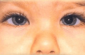
Congenital Glaucoma:
This is the early stage of the disease, therefore having no particular indications. If the eye is not treated, a white lens will cover the eye and the baby will eventually lose eyesight. It is vital that the eye is treated without any delay at the earliest stages of the disease. Glaucoma operations are being done under general anesthesia.
Congenital Cataract:
Cataract operations for old people is nearly the same as with young people. The operation undergone for a Congenital Cataract is applied by PHACO (= FAKO) technical.
Tear Canal Duct:
In most cases, baby’s tear duct will open on its own between six months to one year of age. If it persists past one year operation could be necessary your child’s blocked tear duct does go away, treatments can include.
Nasolacrimal massage, in which you massage the inside corner of your child’s nose 2 to 3 times a day
Cleaning any discharge or matter in the eyes with a warm washcloth
Antibiotic eye drops when the discharge in the eyes becomes excessive, like if you are having to wipe it away more than 2 or 3 times a day
Oral antibiotics if your child develops symptoms of dacryocystitis
Surgical Treatments ; A surgical probe takes about 10 minutes. A thin, blunt metal wire is gently passed through the tear duct to open any obstruction. Sterile saline is then irrigated through the duct into the nose to make sure that there is now an open path. There’s very little discomfort after the probing. If surgical probing is unsuccessful, your doctor may recommend further surgical treatment.
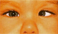
Strabismus:
There are six muscles that control the muscular movement by joining the outer part of each eye. To be able to focus on a certain target by maintaining the eyes parallel, all muscles should function together and in a balance. In strabismus, first the refractive error should be corrected with eye-glasses. In addition, if there is laziness, this should be definitely treated through several methods.Sometimes, these can be sufficient to correct the strabismus.
The cross-eye which cannot be corrected with eye-glasses after the treatment of the amblyopia is treated surgically. Sometimes there is a resistance against the amblyopia treatment, in these cases closing treatment can be performed after the application of the surgery.
The common types of strabismus are :
Turning inward (esotropia): This is the strabismus type most commonly seen at infants. Children with eyes turning inward cannot use both eyes.Mostly, the eyes can be made parallel through surgical procedure in the early period. During the surgical treatment of the esotropia, the tension of one or two muscles is adjusted and the inner muscles are removed and joined to a backward location.
Adaptive turning inward: It is more commonly seen in two-year old or older children. The child can adjust his eyes for near-sight, however, this attempt for focusing causes crossing in the eyes. To make the eyes parallel, eye-glasses, eye drops or special lenses called prism can be applied.
Turning outward (exotropia): This is observed more commonly in children with myopia. An increase in the degree of turning can be seen while looking at the distant objects. It can be usually treated by eye-glasses or strabismus surgery.
Squint Operations:
Squint operations are performed under general anaesthesia. The eye surgeon moves the muscles connected to the eye so that they are strengthened or weakened. This stops the muscles pulling the eye out of alignment. The number of muscles the surgeon moves depends on the type of squint your child has.
Before After
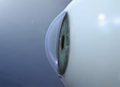
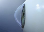
- Keratoconus
This is a connective tissue disease characterized with cornea getting tampered & thin.Since keratoconus is a progressive disease, these complaints tend to gradually grow.In advanced ages, a permanent white spot occurs in the center of the cornea, so keratoplasty (cornea transplantation) is inevitable in this period.
Keratoconus is the thinning of the central zone of the cornea, the front surface of the eye. As a result of this thinning, the normally round shape of the cornea is distorted and a cone-like bulge develops, resulting in significant visual impairment. Keratoconus generally progresses between the ages of 20-40.
Keratoconus usually affects both eyes, though symptoms in each eye may differ. Symptoms usually start to occur in people who are in their late teens and early 20s and may include:
Symptoms
Continuously varying and increasing eye-glasses degrees
Progressing myopiaand astigmatic
Disability to see clearly even when using eye-glasses. Different eye-glasses prescribed by different ophthalmologists
Allergy
Burning sensation, stinging pain, itching and rubbing
Blurred light vision
Double vision
For patients with slight vision in the early period, irregular astigmatism considerably affects the life.It is not possible to treat this type of astigmatic by means of eye-glasses. Because of that keratoconus patients are not satisfied with their eye-glasses prescribed many times or repeating contact lens trials can fail.
Normal Eye The eye with Keratoconus
Treatment:
For patients with slight vision in the early period, irregular astigmatism considerably affects the life.It is not possible to treat this type of astigmatic by means of eye-glasses. Because of that keratoconus patients are not satisfied with their eye-glasses prescribed many times or repeating contact lens trials can fail.
Ring Application ine the Cornea:
It is applied if the keratoconus is not in the advanced period. The cornea ring is placed in the channels that are opened with laser in the cornea. The surgery is made under local anaesthesia. There 3 kinds of intraocular rings in our member hospitals. (Intacs, ferrara, kerraring)
UV Cross Linking (CCL) Treatment:
It is applied in special centers in the world today. The aim of CCL is to stop the thinning of the cornea in the keratoconus. A special operation is applied to the epithelial tissue in the eye, and UVA light and a special medicine called Riboflavin is given. This method is not made for once, it can be repeated. It’s effect on the cornea can be observed as from the 2nd month. It is applied under local anaesthesia.
Cornea Transplant:
Cornea Transplant surgery involves the replacement of the front transparent layer of the eye that is deformed, as a result of some diseases, with healthy cornea tissue. In the cornea transplant, a round section with a diameter of 6-9 mm is removed from the donated healthy cornea, and a part of the cornea of the same size is removed from the patient, and the healthy cornea section is placed in this section. The surgery is performed under general anaesthesia.
- Strabismus
It is referred to as the impairment of the parallelisms in the optic axes of two eyes when they look at a body located at a far distance. Provided that the diagnosis is not established early, it is not possible to treat in the advanced age. So, children should be inspected in the infancy and then at least once in a year.
There are six muscles that control the muscular movement by joining the outer part of each eye. To be able to focus on a certain target by maintaining the eyes parallel, all muscles should function together and in a balance. In strabismus, first the refractive error should be corrected with eye-glasses. In addition, if there is laziness, this should be definitely treated through several methods.Sometimes, these can be sufficient to correct the strabismus.
The cross-eye which cannot be corrected with eye-glasses after the treatment of the amblyopia is treated surgically. Sometimes there is a resistance against the amblyopia treatment, in these cases closing treatment can be performed after the application of the surgery.
Types
Turning inward (esotropia): This is the strabismus type most commonly seen at infants. Children with eyes turning inward cannot use both eyes.Mostly, the eyes can be made parallel through surgical procedure in the early period. During the surgical treatment of the esotropia, the tension of one or two muscles is adjusted and the inner muscles are removed and joined to a backward location.
Adaptive turning inward: It is more commonly seen in two-year old or older children. The child can adjust his eyes for near-sight, however, this attempt for focusing causes crossing in the eyes. To make the eyes parallel, eye-glasses, eye drops or special lenses called prism can be applied.
Turning outward (exotropia): This is observed more commonly in children with myopia. An increase in the degree of turning can be seen while looking at the distant objects. It can be usually treated by eye-glasses or strabismus surgery.
- Uveits
The name of this disease is taken from the inflammation of the eye layer referred to as uvea which consists of iris, choroid and ciliary.Uvea comprises the vessels that nourish the eyes.The inflammation of this layer threats the vision by affecting all tissues of the eye. Uveitis disease is a health problem which can be repeated with long terms, and which can lead to permanent reduction in vision if the correct diagnosis is not established the convenient treatment is not applied.
Primary symptoms of uveitis that can be detected only through the examination in some cases are as follows:
Severe eye pain
Serious blurred vision
Dazzling, sensitivity to light
Eye Redness
Lacrimation
Spots occurring in the field of vision
Uveitis is an urgent disease regardless of the severity and should be intervened immediately. In case of late diagnosis, the disease progresses and may have permanent side effects such as deformities, cataract, high glaucoma, etc. on the pupil. This disease may be diagnosed immediately due to its typical appearance. If the back side of the eye is infected, techniques such as angiography, ultrasonography and ERG in order to determine to what extent the visual ability is threatened and to follow up the efficiency of the treatment may be required. For example, in suspicious cases, angiographies which are performed with a pigment called ICG (indocyanine green) may provide information about the disease which directly leads to diagnosis. After symptoms occur, you should be examined by your ophthalmologist. Uveitis may result in permanent sight impairment.
Although no connection could be found in many cases, viruses, fungi and parasites can cause uveitis for some patients.Furthermore, arthritis located in other parts of the body and diseases like Behçet can trigger uveitis.The most commonly referred treatment for uveitis is still steroid treatment and it is administrated in the form of tablet or in the form intraocular injection.
Behçet’s Disease has three primary symptoms, namely canker sores in the mouth, uveitis in the eye and wounds in genital organs. It is a major vasculitis that may infect various systems such as the skin, joints, digestive and nervous system and large veins. Mediterranean countries, which we call the Silk Road Zone, such as Turkey, Israel and Japan are the ones where this disease is commonly observed. As it is seen, genetic factors are predominant. If not treated, this disease may result in blindness in 2 or 3 years. Since the disease is better understood and more advanced treatment methods are used today, 80% success is achieved.
- Retinal Diseases
Covering the back wall of the eye socket, the network layer consisting of millions of vision cells transmits the images to the optic nerve by means of the nerve fibers. The diseases occurring in that region affect the visual sense negatively. The signs of retinal diseases are : sudden loss of vision, photopsy, the feeling of floating specks, breaking view of the image. Early diagnosis and proper surgical intervention are essential.
Retinal Diseases are retina vascular diseases, diabetes-related diseases, retinal vascular occlusions, retinal detachment, and other age-related macula (yellow spot) disease, congenital retinal disease, intraretinal & under-retina hemorrhages, retinitis pigmentosa, the accumulation of fluid under the retina, retinal vascular diseases, congenital, hereditary diseases & retinal tumors.
Treatment
Retinal Surgery, For large detachments and diabetes-, trauma- and infection-related retinal diseases, usually vitrectomy is applied.
Intraocular injection or photodynamic treatment is applied according to the circumstance in macula detachment treatment. In the treatment of macular degeneration related with age, intraocular injection and/or photodynamic therapy is applied. In photodynamic treatment (FDT), a special substance is injected through the arm. Later on, special laser beams are administered to this pigment which accumulates in malicious veins for a certain period of time with the help of a lens placed in the eye. While malicious veins are destroyed, retina and healthy veins are not damaged. FDT may be repeated if necessary.
Argon laser sticks the retina nerve layer to the pigment layer on it in the region that it is applied and hence the retinal detachment is prevented as a result of the fluid leakage around the laceration. The surgery is applied in the eyes with retinal detachment under the general or local anesthesia. Depending on the level of detachment, detachment surgery from outside of the eye (circlage) or vitrectomy can be applied. In the detachment surgery, a band or a silicon piece is placed on the outer side of the eye.
By means of the vitrectomy surgery, vitreus causing the laceration is removed by entering into the eye, retina is stuck, laser is applied around it and special gases or silicon oil is given into the eye to prevent it to open again.
In detachments with giant laceration and in the tractional detachments developed in the advanced stages of the diabetic retinopathy, in trauma, in the retina damages associated with intraocular foreign body, usually vitrectomy is applied.
- Glaucoma
Glaucoma is a group of diseases that can damage the eye’s optic nerve and result in vision loss and blindness. Glaucoma occurs when the normal fluid pressure inside the eyes slowly rises. However, with early treatment, you can often protect your eyes against serious vision loss.
Risks and diagnosis
Since it is a chronic disease, lifelong glaucoma should be closely supervised by an efficient physician. In that case, blindness risk is very low. It occurs in 1 out of each 40 people over the age of 40.
Eye redness, eye pain, blurred vision, seeing colored rings around lights, nausea and vomiting are the factors that increase the risk of glaucoma with symptoms such as:
Advanced age
Genetic disposition
Smoking
Diabetes
Higher/lower blood pressure
Myopia
Long-term cortisone therapy
Eye injuriest
No symptoms are visible when you have glaucoma. In an advanced stage it will form a threat for our eye-sight. With regular and detailed eye examinations glaucoma can be discovered at the early stages. With NFA (Nerve Fiber Analyzer) are able to determine the damage to the optical nerve fibers. Top determines damage to the nerve fibers at the optical nerve head and the retinal surface.
Treatment
Glaucoma can be treated with eye drops, pills, laser surgery, traditional surgery or a combination of these methods. The goal of any treatment is to prevent loss of vision, as vision loss from glaucoma is irreversible. The good news is that glaucoma can be managed if detected early, and that with medical and/or surgical treatment, most people with glaucoma will not lose their sight.
Taking medications regularly, as prescribed, is crucial to preventing vision-threatening damage. That is why it is important for you to discuss side effects with your doctor. While every drug has some potential side effects, it is important to note that many patients experience no side effects at all. You and your doctor need to work as a team in the battle against glaucoma.
They include:
Eye Drops
It is important to take your medications regularly and exactly as prescribed if you are to control your eye pressure. Since eye drops are absorbed into the bloodstream, tell your doctor about all medications you are currently taking. Ask your doctor and/or pharmacist if the medications you are taking together are safe. Some drugs can be dangerous when mixed with other medications. To minimize absorption into the bloodstream and maximize the amount of drug absorbed in the eye, close your eye for one to two minutes after administering the drops and press your index finger lightly against the inferior nasal corner of your eyelid to close the tear duct which drains into the nose. While almost all eye drops may cause an uncomfortable burning or stinging sensation at first, the discomfort should last for only a few seconds.
Pills
Sometimes, when eye drops don’t sufficiently control IOP ( IntraOcular eye Pressure ), pills may be prescribed in addition to drops. These pills, which have more systemic side effects than drops, also serve to turn down the eye’s faucet and lessen the production of fluid. These medications are usually taken from two to four times daily. It is important to share this information with all your other doctors so they can prescribe medications for you which will not cause potentially dangerous interactions.
Surgical Procedures
When medications do not achieve the desired results, or have intolerable side effects, your ophthalmologist may suggest surgery.
Laser Surgery
Laser surgery has become increasingly popular as an intermediate step between drugs and traditional surgery though the long-term success rates are variable.
The most common type performed for open-angle glaucoma is called trabeculoplasty. This procedure takes between 10 and 15 minutes, is painless, and can be performed in either a doctor’s office or an outpatient facility. The laser beam (a high energy light beam) is focused upon the eye’s drain. Contrary to what many people think, the laser does not bum a hole through the eye. Instead, the eye’s drainage system is changed in very subtle ways so that aqueous fluid is able to pass more easily out of the drain, thus lowering IOP (intraocular eye pressure).
You may go home and resume your normal activities following surgery. Your doctor will likely check your IOP (intraocular eye pressure) one to two hours following laser surgery. After this procedure, many patients respond well enough to be able to avoid or delay surgery. While it may take a few weeks to see the full pressure-lowering effect of this procedure, during which time you may have to continue taking your medications, many patients are eventually able to discontinue some of their medications. This, however, is not true in all cases. Your doctor is the best judge of determining whether or not you will still need medication. Complications from laser are minimal, which is why this procedure has become increasingly popular and some centers are recommending the use of laser before drops in some patients.
Argon Laser Trabeculoplasty (ALT) — for open-angle glaucoma
The laser treats the trabecular meshwork of the eye, increasing the drainage outflow, thereby lowering the IOP. In many cases, medication will still be needed. Usually, half the trabecular meshwork is treated first. If necessary, the other half can be treated as a separate procedure.
This method decreases the risk of increased pressure following surgery. Argon laser trabeculoplasty has successfully lowered eye pressure in up to 75 percent of patients treated. This type of laser can be performed only two to three times in each eye over a lifetime.
Selective Laser Trabeculoplasty (SLT) — for open-angle glaucoma
SLT is a newer laser that uses very low levels of energy. It is termed “selective” since it leaves portions of the trabecular meshwork intact. For this reason, it is believed that SLT, unlike other types of laser surgery, may be safely repeated. Some authors have reported that a second repeat application of SLT or SLT after prior ALT is effective at lowering IOP.
Laser Peripheral Iridotomy (LPI) — for angle-closure glaucoma
This procedure is used to make an opening through the iris, allowing aqueous fluid to flow from behind the iris directly to the anterior chamber of the eye. This allows the fluid to bypass its normal route. LPI is the preferred method for managing a wide variety of angle-closure glaucomas that have some degree of pupillary blockage. This laser is most often used to treat an anatomically narrow angle and prevent angle-closure glaucoma attacks.
Cycloablation
Two laser procedures for open-angle glaucoma involve reducing the amount of aqueous humor in the eye by destroying part of the ciliary body, which produces the fluid. These treatments are usually reserved for use in eyes that either have elevated IOP after having failed other more traditional treatments, including filtering surgery, or those in which filtering surgery is not possible or advisable due to the shape or other features of the eye. Transscleral cyclophotocoagulation uses a laser to direct energy through the outer sclera of the eye to reach and destroy portions of the ciliary processes, without causing damage to the overlying tissues. With endoscopic cyclophotocoagulation (ECP), the instrument is placed inside the eye through a surgical incision, so that the laser energy is applied directly to the ciliary body tissue.
Traditional Surgery
Trabeculectomy
When medications and laser therapies do not adequately lower eye pressure, doctors may recommend conventional surgery. The most common of these operations is called a trabeculectomy, which is used in both open-angle and closed-angle glaucomas. In this procedure, the surgeon creates a passage in the sclera (the white part of the eye) for draining excess eye fluid. A flap is created that allows fluid to escape, but which does not deflate the eyeball. A small bubble of fluid called a “bleb” often forms over the opening on the surface of the eye, which is a sign that fluid is draining out into the space between the sclera and conjunctiva. Occasionally, the surgically created drainage hole begins to close and the IOP rises again. This happens because the body tries to heal the new opening, as if it was an injury. Many surgeons perform trabeculectomy with an anti-fibrotic agent that is placed on the eye during surgery and reduces such scarring during the healing period. The most common anti-fibrotic agent is Mitomycin-C. Another is 5-Fluorouracil, or 5-FU.
About 50 percent of patients no longer require glaucoma medications after surgery for a significant length of time. Thirty-five to 40 percent of those who still need medication have better control of their IOP. A trabeculectomy is usually an outpatient procedure. The number of post-operative visits to the doctor varies, and some activities, such as driving, reading, bending and heavy lifting must be limited for two to four weeks after surgery.
Drainage Implant Surgery
Several different devices have been developed to aid the drainage of aqueous humor out of the anterior chamber and lower IOP. All of these drainage devices share a similar design which consists of a small silicone tube that extends into the anterior chamber of the eye. The tube is connected to one or more plates, which are sutured to the surface of the eye, usually not visible. Fluid is collected on the plate and then absorbed by the tissues in the eye. This type of surgery is thought to lower IOP less than trabeculectomy but is preferred in patients whose IOP cannot be controlled with traditional surgery or who have previous scarring.
Nonpenetrating Surgery
Newer nonpenetrating glaucoma surgery, which does not enter the anterior chamber of the eye, shows great promise in minimizing postoperative complications and lowering the risk for infection. However, such surgery often requires a greater surgical acument and generally does not lower IOP as much as trabeculectomy. Furthermore, long term studies are needed to assess these procedures and to determine their role in the clinical management of glaucoma patients.
Some Promising Surgical Alternatives
The ExPress mini glaucoma shunt is a stainless steel device that is inserted into the anterior chamber of the eye and placed under a scleral flap. It lowers IOP by diverting aqueous humor from the anterior chamber. The ExPress offers the glaucoma surgeon an alternative to either repeating a trabeculectomy or placing a more extensive silicone tube shunt in those patients whose IOP is higher than the optic nerve can tolerate.
The Trabectome is a new probe-like device that is inserted into the anterior chamber through the cornea. The procedure uses a small probe that opens the eye’s drainage system through a tiny incision and delivers thermal energy to the trabecular meshwork, reducing resistance to outflow of aqueous humor and, as a result, lowering IOP.
Canaloplasty, a recent advancement in non-penetrating surgery, is designed to improve the aqueous circulation through the trabecular outflow process, thereby reducing IOP. Unlike traditional trabeculectomy, which creates a small hole in the eye to allow fluid to drain out, canaloplasty has been compared to an ocular version of angioplasty, in which the physician uses an extremely fine catheter to clear the drainage canal.